Here we explain the causes, symptoms, types and diagnosis of vein disorder.
The cause of Vein Disorder
The trigger for vein disorder is often a genetic weakness of the veins. This weakness results from the natural human upright posture. Almost 7000 litres of blood must be pumped through veins against gravity from the feet to the heart. When venous valves are impaired in the case of venous disease, there is a delay in the transport of the blood back to the heart. This at first manifests itself in heavy legs and in a tendency of the legs to swell, and can indicate a wide variety of different health conditions.
In the deep veins
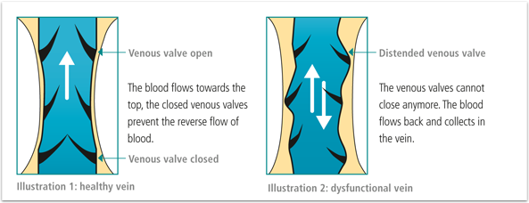
A prerequisite for efficiently functioning venous valves is a venous wall with sufficient tension. Only if the latter requirement is met can the venous valves close properly. If this is not the case - i.e. the venous walls are distended, warped or have lost their shape, e.g. in the case of varicose veins - the distance between the two valves grows and they can no longer close properly. Blood can flow back unhindered. This is called venous insufficiency. The venous blood flows as a consequence from the deep leg veins against its natural direction into the superficial veins and causes blood congestion there. The veins must collect the increased volume of blood and expand as a result.
In the superficial veins
If the venous valves of the superficial venous system are defective, the deep venous system has to transport more blood than usual. Blood volume therefore increases and the veins distend.
Result: The venous valves can no longer close properly and the blood is only directed back to the heart with delay.
This at first manifests itself in heavy legs and in a tendency for the legs to swell, and can reflect a range of different types of disease, such as varicosis (varicose veins) and complications following thromboses. The associated discomfort and changes to the skin of the legs are referred to as chronic venous insufficiency (CVI).
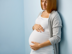
Pregnancy can be a cause of vein disorder.
These factors favour the development of varicose veins
Impaired blood flow:
clothing that is too tight and pinches
crossed legs
blood volume too high as a result of pregnancy
sitting or standing for long periods of time
unhealthy diet, obesity
Relaxed venous wall:
alcohol
heat
hormones (contraceptive pill, hormones against menopause, pregnancy)
Impaired muscle pump:
wrong shoes (high heels)
sitting or standing for long periods of time
paralysis
Heridity:
congenital weakness of connective tissue and the venous walls
Types of venous disease
A tendency towards venous disease is hereditary, while hormonal influence (pregnancy, the pill), activities that require mainly sitting or standing for long periods of time, obesity and bad shoes demonstrably favour the incidence of vein problems. In Germany, more than 50% of the population suffer at least a slight change of the venous system. Early diagnosis and rigorous treatment can alleviate existing complications and protect the patient against serious subsequent damage. However, if they remain untreated, they can cause serious suffering.
In Germany, more than 50% of the population suffer at least a slight change of the venous system.
Spider veins
Spider veins are small, red or bluish looking veins, also called micro varicose veins, that lie just below the skin's surface. They are a few millimetres or centimetres long, have a diameter of up to one millimetre and are often arranged in a fan-shaped structure. Spider veins, in contrast to varicose veins, are not a disease but usually only an aesthetic-cosmetic problem that can be corrected using a range of treatment options. However, they can also be a warning signal for varicose veins present just below. A visit to a physician can clarify the cause.
Varicose veins
A varicose vein (varix) is an extended, wildly weaving vein in the skin. It develops when the wall of the veins is distended so much that the venous valves can no longer close and the blood congests. This results in so-called “phlebostasis”. In comparison with harmless spider veins, varicose veins are a real disease that without the correct treatment may have serious consequences, such as inflammation of the veins, chronically venous insufficiency, vascular occlusion, etc. Timely therapy is therefore enormously important.
Thrombosis
Thrombosis develops when a blood clot (a thrombus) forms on the vascular wall. This obstructs the veins, the venous valves no longer function and this prevents the blood from flowing back. The result is blood congestion. Most blood clots develop in the deep veins of the leg and pelvis and become apparent through sudden pain in the calves or swelling or a heavy feeling in the legs. A blood clot can develop into a fatal pulmonary embolism. That is why it is absolutely essential to visit a physician at the first signs of symptoms. Risk factors are sitting for long periods of time (above all while travelling), varicose veins and an increased tendency towards blood clots following an operation or birth of a child.
Pulmonary embolism
A pulmonary embolism may occur as a result of thrombosis. Here the blood clot detaches itself and is released into the bloodstream and carried right into the minute branches of blood vessels in the lungs. It may happen that the blood clot is lodged there and disturbs the oxygen supply of the lungs. The result is a pulmonary embolism. The lungs can no longer fully perform their original function. In some cases, pulmonary embolism can lead to death.
Inflammation of the vein (phlebitis)
A frequent complication of pronounced varicose veins is the inflammation of a venous vessel (phlebitis). It is felt and seen as a painful, bright red and heated strand in the course of the varicose vein often accompanied by swelling. The causes are the blood clots in the superficial vein resulting from the inflammation.
An inflammation of the vein must receive immediate medical treatment, as it could otherwise grow and the blood clots could be embedded in the deep vein system. Untreated varicose veins can develop into chronically venous insufficiency with potential swelling and changes to the skin, and even leg ulcers.
Open legs
Long-lasting venous congestion may lead as a result of the undersupply of the affected tissue to devastating damage of the cells and to tissue necrosis. The result is long lasting open wounds.
Diagnosis
Vein diagnostics has several methods to determine the type and severity of the venous disease. The goal of all of these measures is to find a therapy that is fine-tuned to the individual. The following methods are common:
Anamnesis
In the case of anamnesis, the physician establishes a patient’s medical history with regard to their current ailment. With venous problems, the patient is asked about earlier thromboses and inflammation of the veins. The physician further determines any tendency for water to collect in the legs. Women are also asked about pregnancy or whether they take hormonal contraception (the contraceptive pill). And one’s family history is of relevance: family-related disease patterns provide information about a possible tendency towards varicose veins and vein problems.
This is then followed by the physician closely examining the legs in different body positions. This is because varicose veins fill up totally when standing, and this is not the case when lying down. The skin, especially the skin around the ankle, is also checked very thoroughly. Should edema (water retention in tissue) be suspected, the physician will press the swollen leg. If an indentation occurs that only disappears slowly, this indicates an edema.
Ultrasonic testing
Different kinds of equipment, primarily the Doppler ultra-sonography (a special kind of ultrasonic testing), are available to determine the patient’s medical condition. This method allows for the speed and direction of blood flow to be measured and obstructions or defective venous valves to be detected. By means of duplex and colour duplex sonography, an additional ultrasound image of the blood vessels can be generated, which can provide more detailed information about the state and capability of the veins. This method is always used when thrombosis or inflammation of the veins is suspected, when other methods have not provided a clear image or when an operation is planned.
Light-reflection rheography
Photoplethysmography (also called light-reflection rheography, in short LRR) can be additionally used to determine the function of the venous system. The skin of the lower leg is irradiated in a specific area with infrared light and measurements are taken to determine how strongly the light is reflected. As the amount of reflected light is dependent on the amount of blood in the veins below, this examination provides evidence of how well the venous system is functioning.
During the examination, the patient has to move their foot several times in a certain way, as a result of which the veins under observation gradually empty. The examiner then measures how long it takes till the veins are filled with blood again. Should it take longer than 25 seconds, everything is fine. Should the veins fill more quickly, this indicates venous insufficiency because the blood flows back through the damaged venous valves against the normal direction of blood flow.
Please note: This article is for informational purposes only and is not a substitute for professional medical advice.